Cell and flare in the eye (Video)
5 (662) · $ 10.00 · In stock
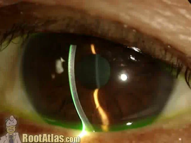
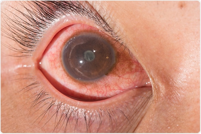
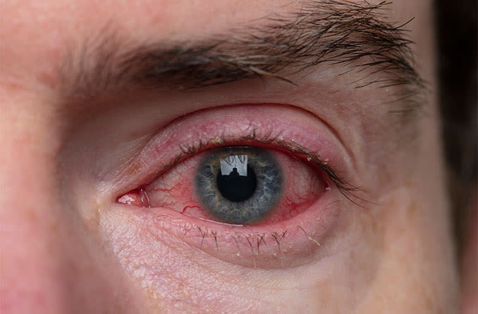
This video demonstrates what cell and flare look like under the slit-lamp microscope. “Cell” is the individual inflammatory cells while “flare” is the foggy appearance given by protein that has leaked from inflamed blood vessels. This finding is commonly seen with uveitis, iritis, and after surgery … and actually seeing it can be challenging for
8,400+ Nerve Cell Stock Videos and Royalty-Free Footage - iStock

Averted vision: how to get a better view of night-sky objects - BBC Sky at Night Magazine
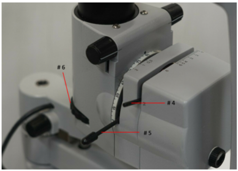
Slit Lamp Basics

Slit Lamp Examination - EyeWiki

Ali Jaworski
Iris Beam's Instagram, Twitter & Facebook on IDCrawl

Block 7: Biomicroscopy Illumination Techniques Flashcards

Woche 10 - mariaundleaunterwegss Webseite!
Blepharitis (Eyelid Inflammation): Symptoms, Causes and Treatment

South Alabama Emergency Medicine (@SouthEmergency) / X

Anterior Chamber Cells