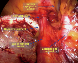
Figure 6 from Femoral Hernia: A Review of the Clinical Anatomy and
4.8 (776) · $ 18.99 · In stock

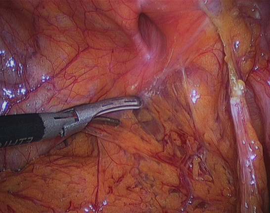
Figure 6. Femoral hernia repair in clean operation. (a) The narrow side of the mesh is sutured to Cooper’s ligament; (b) The mesh is sutured to the iliopubic tract or shelving portion of the inguinal ligament; (c) The posterior wall of the inguinal canal is reinforced, as in Lichtenstein’s repair. - "Femoral Hernia: A Review of the Clinical Anatomy and Surgical Treatment"
Figure 6 from Femoral Hernia: A Review of the Clinical Anatomy and Surgical Treatment
Embryonic developmental process and clinical anatomy of the preperitoneal fascia and its clinical significance

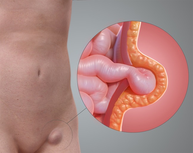
Illustration Of A Femoral Hernia Art Print by John Bavosi - Fine Art America

Femoral Hernia - A Review of Clinical Anatomy

Contemporary management of obturator hernia

Femoral Hernia: A Review of the Clinical Anatomy and Surgical Treatment
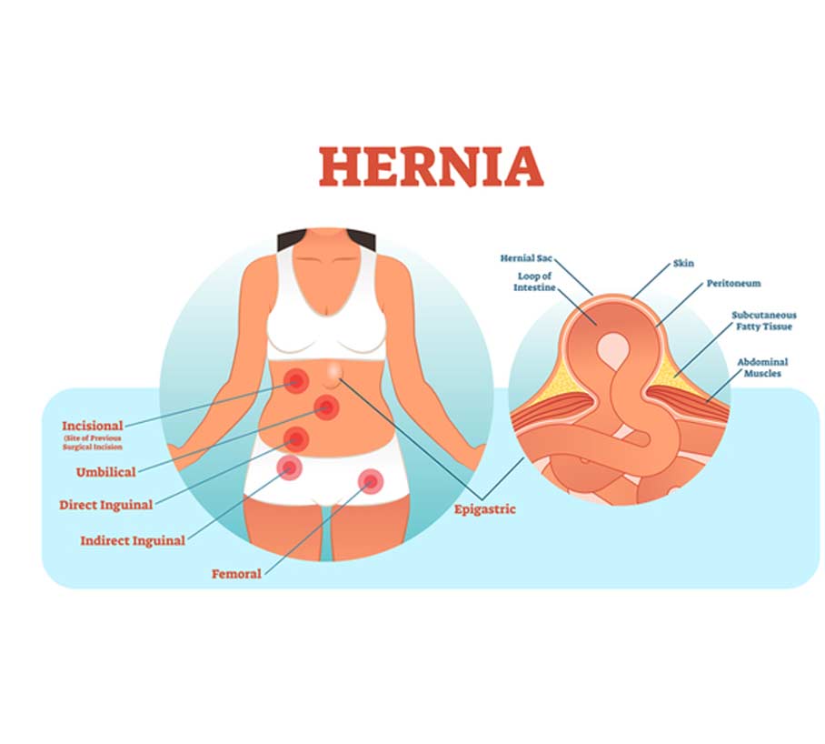
Hernias, Inguinal, Femoral, Umbilical

Clinical Anatomy of the Groin: Posterior Laparoscopic Approach
Femoral hernia