Optical Coherence Tomography: Imaging Mouse Retinal Ganglion Cells In Vivo
4.7 (313) · $ 18.99 · In stock

Scientific Article | Structural changes in the retina are common manifestations of ophthalmic diseases.
Retinal ganglion cell repopulation for vision restoration in optic neuropathy: a roadmap from the RReSTORe Consortium, Molecular Neurodegeneration

PSRC - In Vivo Evaluation Of The Retina And Optic Nerve After Whole Eye Transplantation Using Optical Coherence Tomography, Manganese-enhanced Magnetic Resonance Imaging And Electroretinography

PDF) Modified protocol for in vivo imaging of wild-type mouse retina with customized miniature spectral domain optical coherence tomography (SD-OCT) device

Frontiers Topical nerve growth factor prevents neurodegenerative and vascular stages of diabetic retinopathy

In vivo imaging of mouse retina. The spectral domain-optical
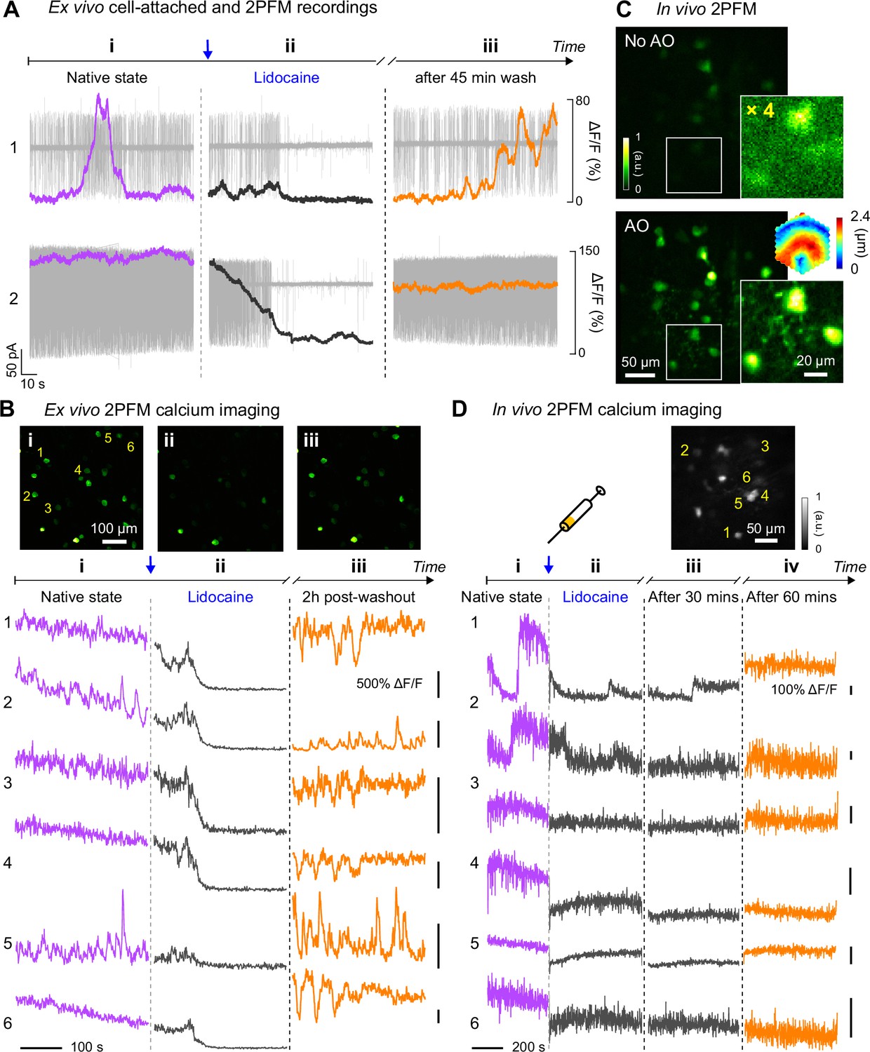
Retinal microvascular and neuronal pathologies probed in vivo by adaptive optical two-photon fluorescence microscopy
Longitudinal In Vivo Imaging of Retinal Ganglion Cells and Retinal Thickness Changes Following Optic Nerve Injury in Mice

Automatic counting of retinal ganglion cells in the entire mouse retina based on improved YOLOv5

All Protocols and Video Articles in JoVE

PDF) Topical nerve growth factor prevents neurodegenerative and vascular stages of diabetic retinopathy

Adaptive optics with combined optical coherence tomography and