- Home
- lumbar compression
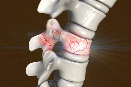
- Lumbar Compression Fracture, Illustration - Stock Image - C027/6314 - Science Photo Library
Lumbar Compression Fracture, Illustration - Stock Image - C027/6314 - Science Photo Library
4.8 (653) · $ 17.50 · In stock

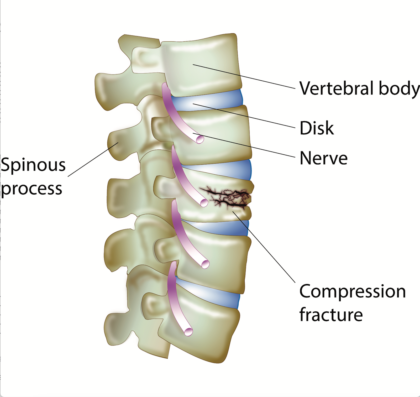
An interpretive illustration of an MRI depicting a sagittal view of compression fractures at the L1 and L2 vertebrae as a result of osteoporosis. Over time as bone becomes weaker and more porous, they become more susceptible to injury and fractures, especially in situations where significant weight or stress is placed on the bone. Evan Oto/SCIENCE PHOTO LIBRARY
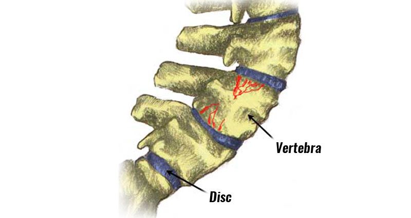
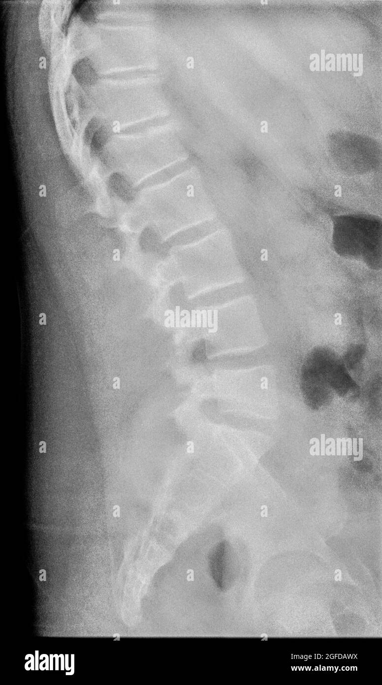
Compression Fracture Of A Lumbar Vertebra #2 Photograph by Zephyr/science Photo Library
Compression Fracture Of A Lumbar Vertebra #2 by Zephyr/science Photo Library

Compression Fracture Images – Browse 2,195 Stock Photos, Vectors, and Video

2,934 Compression Fracture Royalty-Free Photos and Stock Images

Thoracic vertebral collapse, X-ray - Stock Image - C038/6661 - Science Photo Library
Compression fracture spine hi-res stock photography and images - Alamy

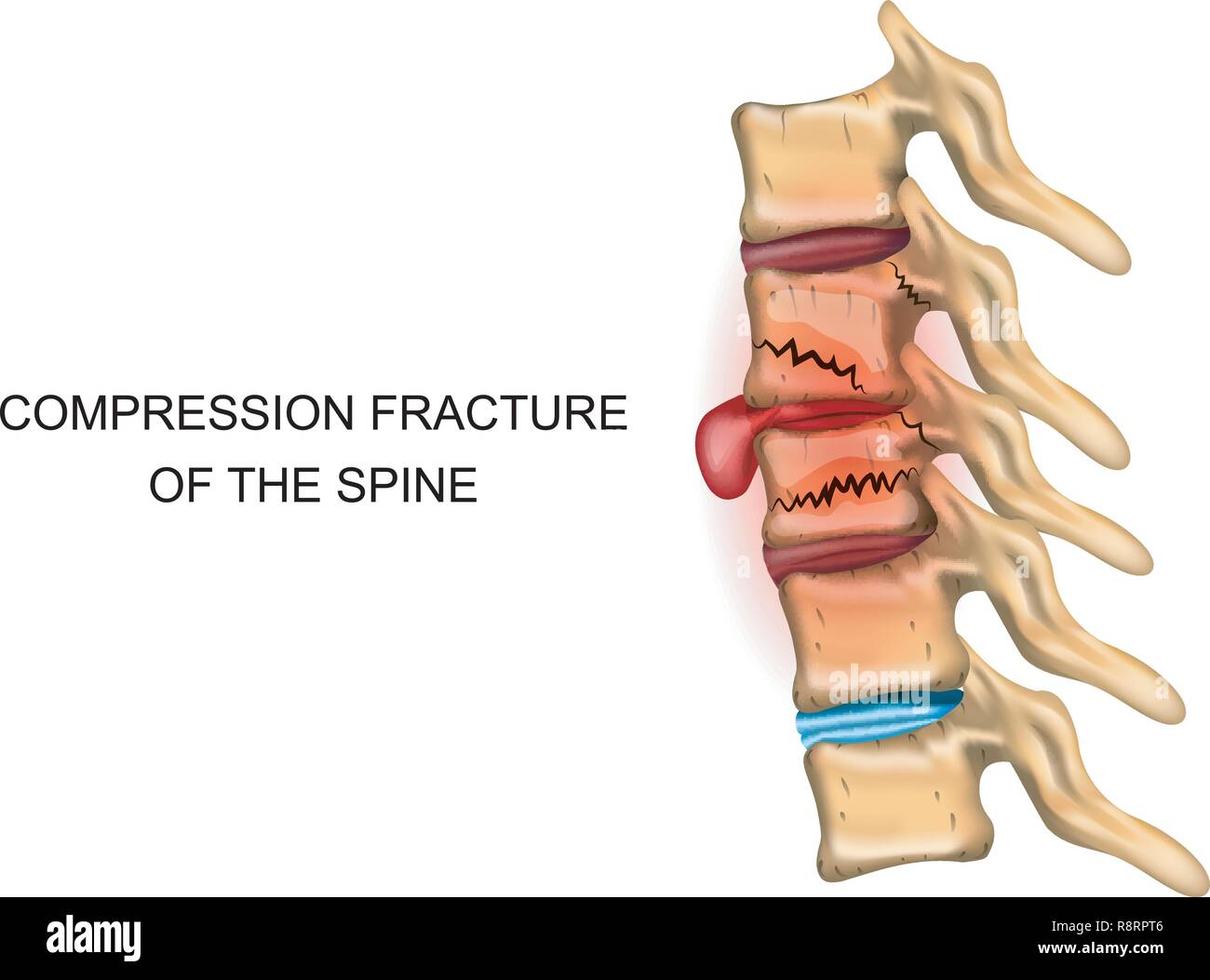
Medical Illustration Symptoms Vertebral Compression Fracture Stock Vector (Royalty Free) 1960120894

lumbar compression fracture - Keyword Search - Science Photo Library

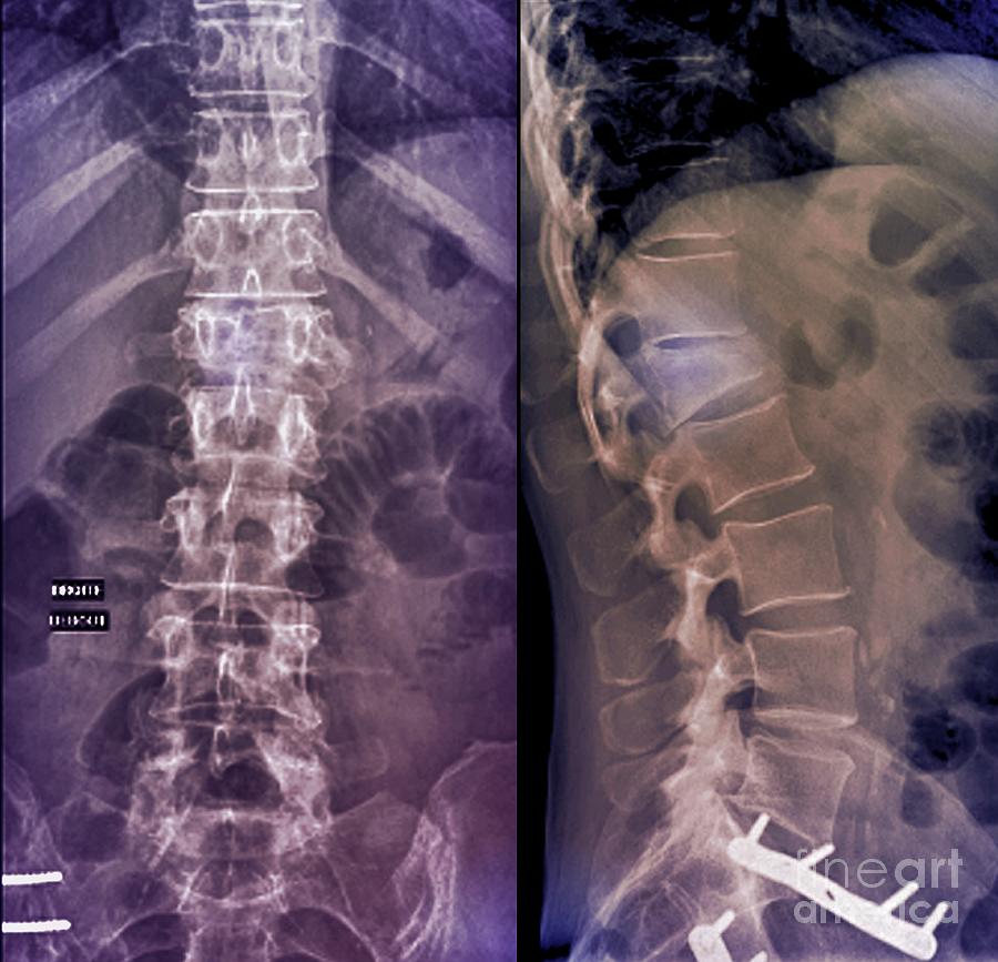
Acute Compression Fracture Of L2 B Metal Print by Living Art Enterprises, LLC - Fine Art America

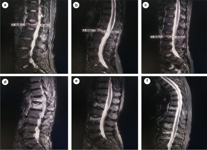
Osteoporosis, MRI scan - Stock Image - C052/9280 - Science Photo Library
Compression fracture spine hi-res stock photography and images - Alamy
Compression Fracture Images – Browse 2,195 Stock Photos, Vectors, and Video
Lumbar Compression Fracture: Practice Essentials, Pathophysiology, Epidemiology