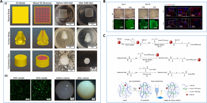
The STL images of two geometries of the 3D-printed bioceramic model
4.6 (791) · $ 22.00 · In stock

Download scientific diagram | The STL images of two geometries of the 3D-printed bioceramic model were designed as follows: The cylindrical compression sample (a), the concave-topped disk structures views of the bottom (c), and the top (d). The cross-section views of concave-top disk structures also showed the STL image of a horizontal section (e) and a vertical section (f). Furthermore, the two kinds of 3D-printed sintered bioceramic images were obtained. The 3D cylinder bioceramic sample (b), the bottom view (g), and the top view (h) of the concave-top disc structure of the 3D-printed bioceramic scaffold from publication: Bilayer osteochondral graft in rabbit xenogeneic transplantation model comprising sintered 3D-printed bioceramic and human adipose-derived stem cells laden biohydrogel | Reconstruction of severe osteochondral defects in articular cartilage and subchondral trabecular bone remains a challenging problem. The well-integrated bilayer osteochondral graft design expects to be guided the chondrogenic and osteogenic differentiation for stem cells and | Bioceramics, Osteochondritis and Grafts | ResearchGate, the professional network for scientists.

ars.els-cdn.com/content/image/1-s2.0-S138589472401
A) Top and crosssectional confocal images of the actin filaments
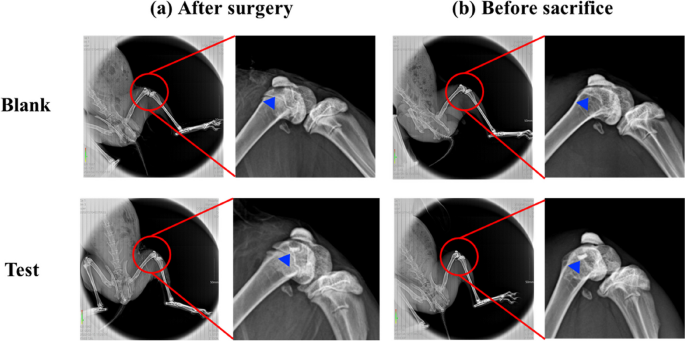
Bilayer osteochondral graft in rabbit xenogeneic transplantation
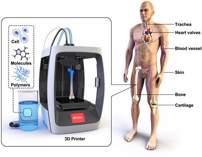
Emerging 3D bioprinting applications in plastic surgery

5776 PDFs Review articles in NANO-SILICA

Shun-Cheng WU, Fellow, Doctor of Philosophy
.JPG)
Leveraging Additive Manufacturing to Improve Joint Replacement

5792 PDFs Review articles in NANO-SILICA
Frontiers Toward Biomimetic Scaffolds for Tissue Engineering: 3D

SEM images of previously microfabricated platforms for bone TE