Ultrasound imaging - Download as a PDF or view online for free
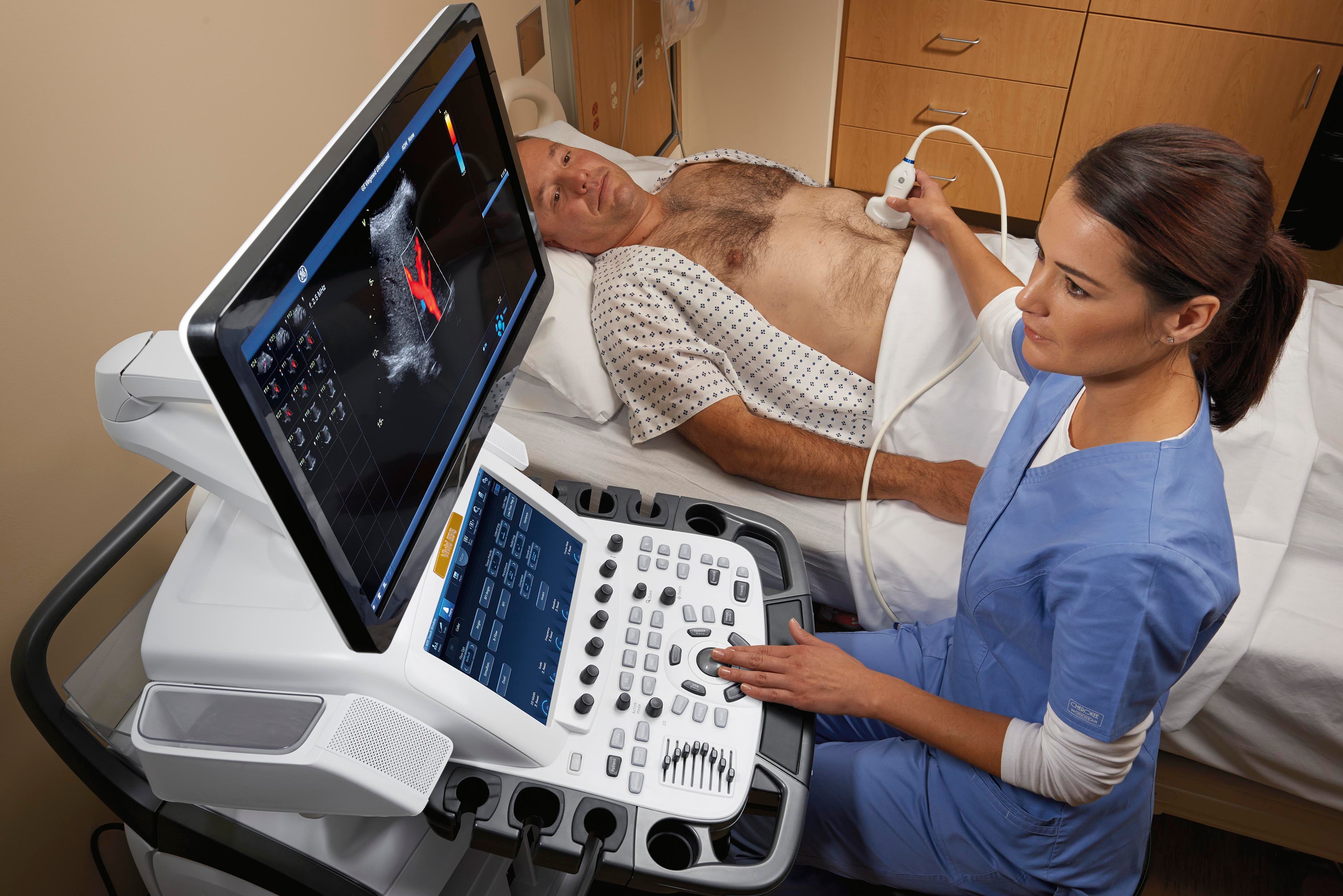
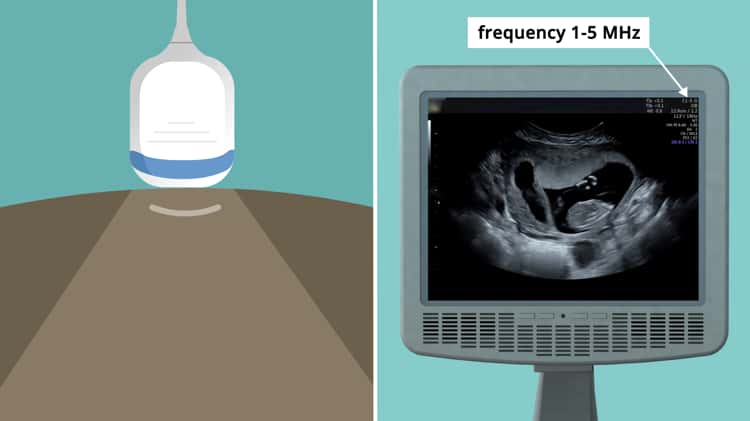
Ultrasound uses high frequency sound waves to visualize internal structures. It works by transmitting sound waves into the body using a transducer probe, which detects the echoes as they bounce off tissues and organs. The echoes are processed to form images on the ultrasound machine screen in real-time. Common applications include obstetrics, cardiology, and urology. The Philips HD11 is an ultrasound system with curvilinear, linear, and phased array probes for different exams. It provides grey scale, Doppler, and color imaging modes. Ultrasound has benefits of being non-invasive, portable, and having no radiation, but has limitations of being operator dependent and unable to penetrate bone.

Acai Ultrasound Imaging Services Ltd.
★𝐌𝐎𝐁𝐈𝐋𝐄 𝐓𝐑𝐎𝐋𝐋𝐄𝐘 𝐂𝐀𝐑𝐓:The ultrasound cart is used for carrying any B-ultrasonic machine and scanner. ★𝐏𝐑𝐄𝐌𝐈𝐔𝐌 𝐌𝐀𝐓𝐄𝐑𝐈𝐀𝐋:It is made of safe material. ABS

Mobile Ultrasound Trolley CartPortable Ultrasound Imaging Scanner Vehicle ABS Plastic 4 Wheels 2 Brake with 3 Holes and Push Handle

Special probes improve ultrasound imaging in obese patients
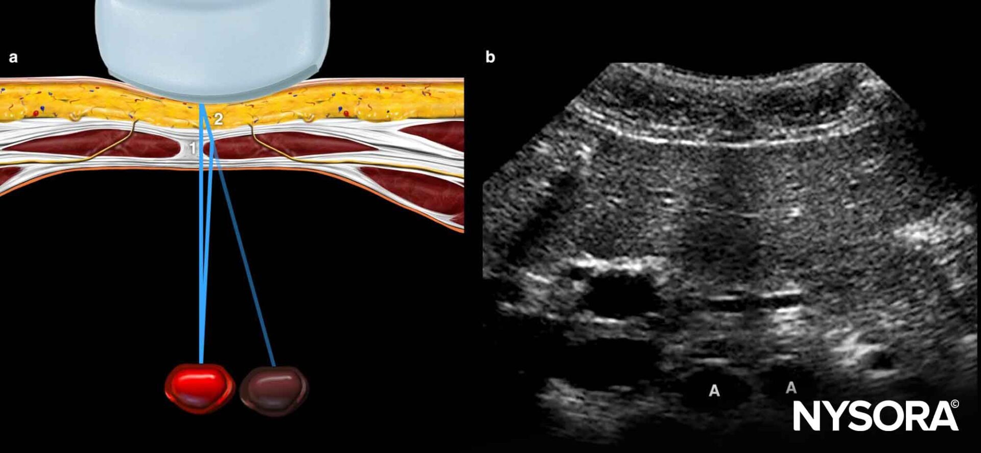
Ultrasound image artifacts explained - NYSORA
Ultrasound - Imaging Healthcare Specialists
Advancements in Ultrasound

Reading Minds with Ultrasound: A Less-Invasive Technique to Decode the Brain's Intentions
The Principles of Ultrasound Imaging on Vimeo
Learning Ultrasound Imaging

AI in Ultrasound Imaging Market Size
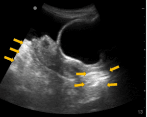
An ultrasound imaging artifact, Case Studies

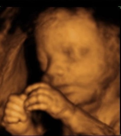
Fetal Pictures of Ultrasounds Gallery
3D Ultrasound - Innovatus Imaging
Highlight, take notes, and search in the book

Radcases Ultrasound Imaging (Radcases Plus Q&A)

Acai Ultrasound Imaging Services Ltd.