Structure of YF1. a Structure of YF1 in its dark-adapted state as
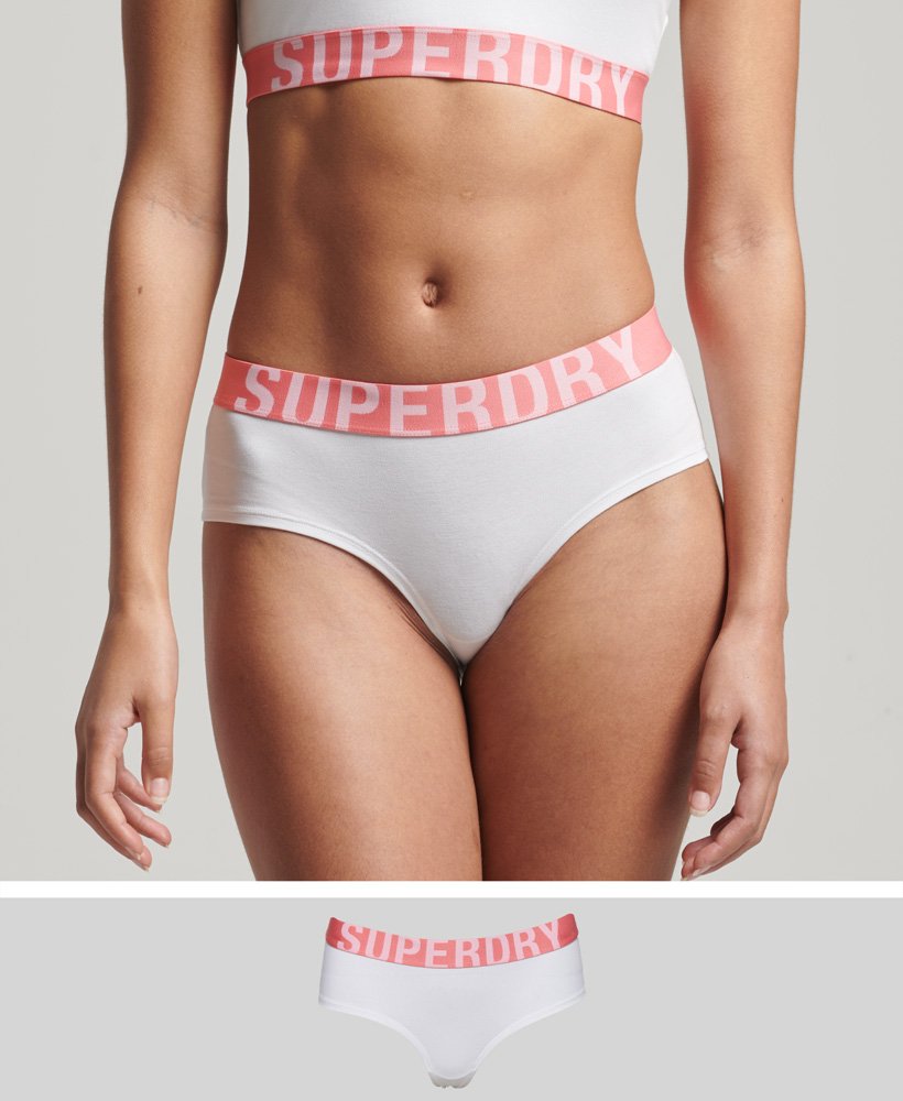
4.9 (396) · $ 22.50 · In stock

Download scientific diagram | Structure of YF1. a Structure of YF1 in its dark-adapted state as resolved by X-ray crystallography 13. The location of the different domains, of the flavin mononucleotide (FMN), of the cofactor adenosine diphosphate (ADP), and of the phosphoaccepting histidine 161 are indicated. b Light induced conformational changes of the LOV photosensor domain refined from X-ray solution scattering 22. The changes are maximal at the C-termini that feed into the Jα helices (dashed arrows). The coloring is according to the root mean square deviation of the alpha carbons from publication: Sequential conformational transitions and α-helical supercoiling regulate a sensor histidine kinase | Sensor histidine kinases are central to sensing in bacteria and in plants. They usually contain sensor, linker, and kinase modules and the structure of many of these components is known. However, it is unclear how the kinase module is structurally regulated. Here, we use | Secondary Protein Structure, Bacterial Proteins and Protein Conformation | ResearchGate, the professional network for scientists.

Cytoskeleton structure of TF1 cells revealed by confocal microscopy.

Stephan NIEBLING, Postdoc, Applied Physics

Light-Oxygen-Voltage (LOV)-sensing Domains: Activation Mechanism and Optogenetic Stimulation - ScienceDirect
IJMS, Free Full-Text
INTERNAL MEMORANDUM TO: Dr Ollivier Dyens, Vice-Provost

Gemma NEWBY, Application Scientist, PhD Chemistry, Xenocs, Sassenage, Science and Application

Gemma NEWBY, Application Scientist, PhD Chemistry, Xenocs, Sassenage, Science and Application

Stephan NIEBLING, Postdoc, Applied Physics

Structure of PAL in its dark-adapted state a, The homodimeric receptor